Welcome to the MRI Lab of the University of Regensburg


Research conducted in our Lab employs a variety of in-vivo imaging techniques to better understand the functions and structures of the human brain in a safe and effective environment

Our group of research is interdisciplinary including scientists from Computer Science, Engineering, Physics, Psychology, Psychiatry and clinical investigators from Neurology, Radiology, and Surgery, as well as students from graduate programs
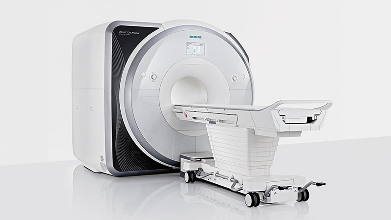


-MRI Acquisition

  • T1 and T2
    • Weighted structural imaging enabling white/grey matter parcellation.
  • FLAIR (FLuid Attenuated Inversion Recovery)
    • T2 weighting with fat saturation to observe cerebellar hyper/hypo intense signals.
  • MRA (Magnetic Resonance Angiography)
    • Visualizing brain vasculature
  • EPI (Echo Planar Imaging)
    • T2* weighted image, used for showing Blood Oxygen Level Dependent (BOLD) signal showing cerebral blood flow in activated regions, at rest or under stimulus.
  • MRS (Magnetic Resonance Spectroscopy)
    • Non imaging technique used to quantify concentrations of metabolites in tissue.
  • Diffusion (HARDI,DTI,DKI)
    • T2 weighted imaging that enables viewing of water molecules diffusion in the brain.
  • SWI (Susceptibility Weighted Imaging)
    • Technique that looks at venous behavior in the brain which can show bleeding and areas of iron deposits.
  • QSM (Quantitative Susceptibility Mapping)
    • QSM utilizes phase images, solves the magnetic field to susceptibility source inverse problem, and generates a three-dimensional susceptibility distribution. Potential QSM applications include standardized quantitative stratification of cerebral microbleeds, neuroinflammation, abnormal iron concentrations and neurodegenerative diseases.


Contact: This email address is being protected from spambots. You need JavaScript enabled to view it.